Medulla spinalis nukleusları [Nuclei of the spinal cord]. Sengul G. 2019. In Fonksiyonun cerrahi anatomisi [Surgical anatomy of the function] Vol 1. Eds. Biceroglu H, Tonge M, Seckin M, Adiguzel E, Gurvit H, Hanci M. pp399-402.
Medulla spinalis bağlantı yolları [Projections of the spinal cord]. Sengul G. 2019. In Fonksiyonun cerrahi anatomisi [Surgical anatomy of the function] Vol 1. Eds. Biceroglu H, Tonge M, Seckin M, Adiguzel E, Gurvit H, Hanci M. pp607-622.
Medulla spinalis fonksiyonel organizasyonu ve mikrocerrahi anatomisi [Functional organization and microsurgical anatomy of the spinal cord]. Sengul G. 2019. In Fonksiyonun cerrahi anatomisi [Surgical anatomy of the function] Vol 2. Eds. Biceroglu H, Tonge M, Seckin M, Adiguzel E, Gurvit H, Hanci M. pp883-890.
Microsurgical anatomy of safe entry zones on the ventrolateral brainstem: a morphometric study. Kocer IB, Demiralin MO, Erturk M, Arslan D, Sengul G. Neurosurg Rev. 2022;45(2):1363-1370. [Link to paper]
Topographic classification of the thalamus surfaces related to microneurosurgery: A white matter fiber microdissection study. Serra C, Türe U, Krayenbühl N, Sengul G, Yaşargil DC, Yaşargil MG. World Neurosurgery 97:438-452, 2017. [Link to paper]
Morphometry of the cervical vertebral pedicles as a guide for transpedicular screw fixation. Kayalioglu G, Erturk M, Varol T, Cezayirli E. Neurol Med Chir 47:102-107, 2007. [Link to paper]
Topographic anatomy of the fornix as a guide for transcallosal-interforniceal approach with a special emphasis on sex differences. Ozer MA, Kayalioglu G, Erturk M. Neurologia Medico-Chirurgica 45:607-612, 2005. [Link to paper]
Variations in sphenoid sinus anatomy with special emphasis on pneumatization and endoscopic anatomic distances. Kayalioglu G, Erturk M, Varol T. Neurosciences 10:79-84, 2005. [Link to paper]
Heights of the cerebral falx. Kayalioglu G, Erturk M, Varol T. Neurosciences 9:257-260, 2005. [Link to paper]
The cranio-orbital foramen, the groove on the lateral wall of the human orbit and the orbital branch of the middle meningeal artery. Erturk M, Kayalioglu G, Govsa F, Varol T, Ozgur T. Clinical Anatomy 18:10-14, 2005. [Link to paper]
Anatomy of the clinoidal region with a special emphasis on the caroticoclinoid foramen and the interclinoid osseous bridge in a recent Turkish population. Erturk M, Kayalioglu G, Govsa F. Neurosurgical Review 27:22-26, 2004. [Link to paper]
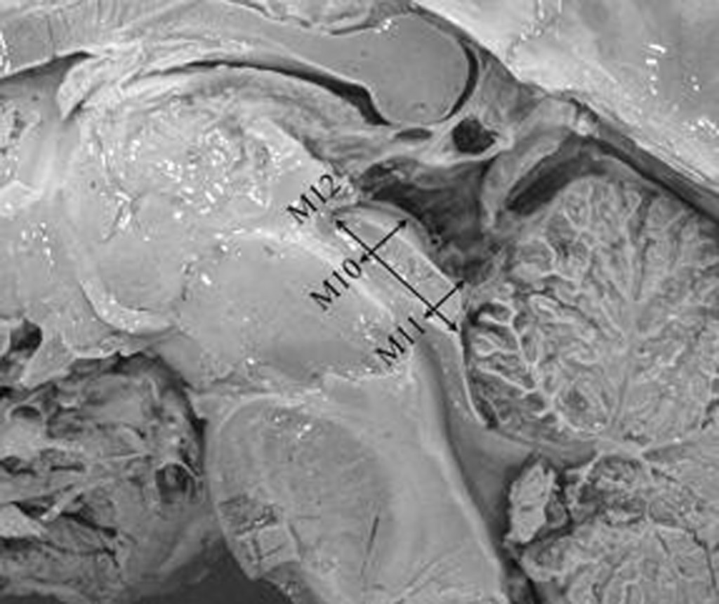
Morphometry of the anterior third ventricle region as a guide for the transcallosal-interforniceal approach. Erturk M, Kayalioglu G, Ozer MA. Neurologia Medico-Chirurgica 44:288-292, 2004. [Link to paper]
Morphometry of the anterior third ventricle region as a guide for the subfrontal (translaminaterminalis) approach. Erturk M, Kayalioglu G, Ozer MA. Neurosurgical Review 26;249-252, 2003. [Link to paper]
Nasal cavity and paranasal sinus bony variations: a computed tomographic study. Kayalioglu G, Oyar O, Govsa F. Rhinology 38:108-113, 2000. [Link to paper]
Neuro-arterial relationships in the region of the optic canal. Govsa F, Erturk M, Kayalioglu G, Pinar Y, Ozer MA. Surgical and Radiologic Anatomy 21:329-335, 1999. [Link to paper]
The cavernous sinus: topographic morphometry of its contents. Kayalioglu G, Govsa F, Erturk M, Pinar Y, Ozer MA, Ozgur T. Surgical and Radiologic Anatomy 21:255-260, 1999. [Link to paper]
The superior orbital fissure and its contents. Govsa F, Kayalioglu G, Erturk M, Ozgur T. Surgical and Radiologic Anatomy 21:181-185, 1999. [Link to paper]
An anatomical study on the sigmoid sulcus and related structures. Kayalioglu G, Govsa F, Erturk M, Arisoy Y, Varol T. Surgical and Radiologic Anatomy 18:289-294, 1996. [Link to paper]
Assessment of normal clivus related to age with magnetic resonance imaging. Oyar O, Govsa F, Sener RN, Kayalioglu G. Surgical and Radiologic Anatomy 18:47-49, 1996. [Link to paper]
